Five studies of the neurovascular unit, all read out of public electron microscopy volumes
rather than collected at a microscope. That is what makes them possible at all here: there is
no laboratory behind this site and no funding — a volume somebody else has already imaged and
released is something one person can work through in their spare time, which is what this is.
Each study below carries what we found, the figure, and the coordinates the figure was taken
at — so you can open the same place in the tools on this site and look for yourself.
Three groups brought the questions, the animals and the physiology, and each of them has a
laboratory. The volume electron microscopy is the half done on this side: one person, a
laptop and a dataset somebody else paid to collect.
Gary P. MorrisTasmanian School of Medicine, University of Tasmania, Hobart
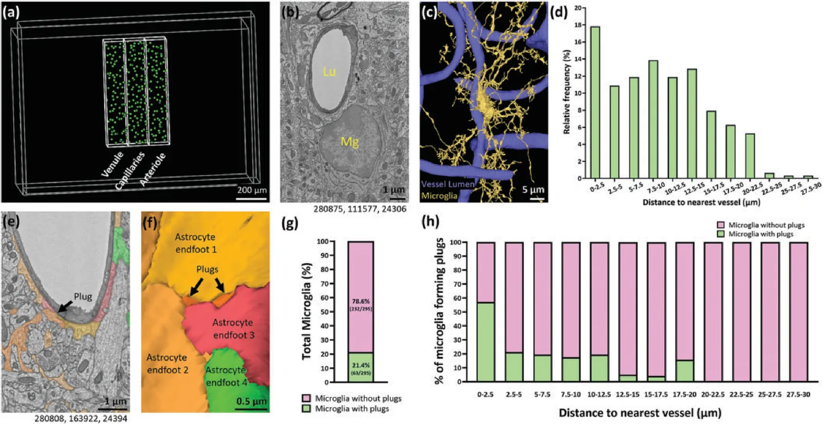
Led the microglia study: 307 microglia annotated one z-plane at a time to ask how often a microglial process reaches the vessel wall through a gap between astrocyte endfeet.
Amreen MughalNeurovascular Research Unit, NINDS & NHLBI, National Institutes of Health, Bethesda
Co-corresponding author on the primary cilia work — pericyte and endothelial cilia and centrioles across mouse and human microvasculature, in three volumes and nine other tissues.
Changsi CaiDepartment of Neuroscience, University of Copenhagen
Corresponding author on the leptomeningeal fibroblast work — two-photon Ca²⁺ imaging in awake mice. The volume EM half, tracing the fibroblast groups over the pial surface, was the contribution from this side.
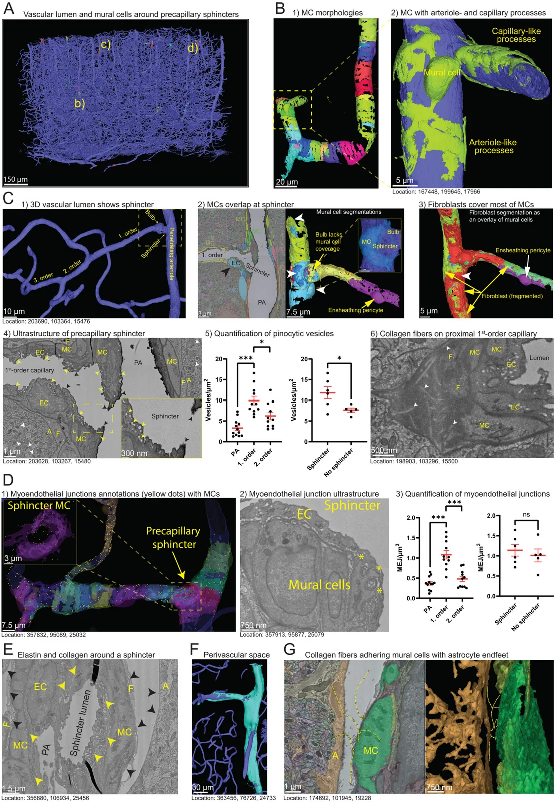
The paper that started the tools. A precapillary sphincter is a ring-shaped narrowing where a penetrating arteriole gives off its first capillary, and almost nothing was known about what it is made of. Reading it in a volume meant every cell of the neurovascular unit could be looked at in the same place at once.
What we found
At the sphincter the mural cell is not a band but several layers of processes wrapped around the indentation — against a single layer on the arteriole above it and on the capillary below.
Endothelial pinocytic vesicles are densest immediately downstream of the sphincter (≈10/µm² on first-order capillaries against ≈3.5/µm² on the arteriole), and denser on branch points that have a sphincter than on those that do not.
Collagen fibrils made by perivascular fibroblasts reinforce the proximal first-order capillary, where the pressure drop is steepest.
Neuronal processes lie perpendicular to the vessel around penetrating arterioles and arterial-end capillaries — in mouse, and again in the human h01 volume — but not around ascending venules.
Figure 1 — the sphincter in seven views, from the whole vascular lumen of the volume down to a 300 nm inset of the indentation itself. Every panel prints the coordinate it was taken at; those are the links below. Figure 1, Grubb 2023, Vascular Biology. CC BY 4.0.
Astrocyte endfeet are usually described as covering the vessel completely. They do not quite: there are gaps between neighbouring endfeet, and the question was whether anything uses them. Three regions of interest were cut through the volume — one around a venule, one around an arteriole, one through the capillary bed between them — and every microglial nucleus in them was annotated by hand.
What we found
Of 307 microglia annotated, 21.4% (63 of 295 scored) touch the vessel directly through a gap between adjacent astrocyte endfeet. We called the contacts plugs.
A plug-forming microglia makes a median of 2 and as many as 17, almost all within a few micrometres of the soma; the closer the cell sits to the vessel, the more it makes.
Plugs are enriched at the venule end of the tree, and contact endothelial cells over pericytes 3:1.
They range from 0.01 µm² to about 15 µm², but 95% are under 1 µm² — small enough that light microscopy would not have separated them from a near miss. 61.8% are bounded by two endfeet and 33.9% by three.
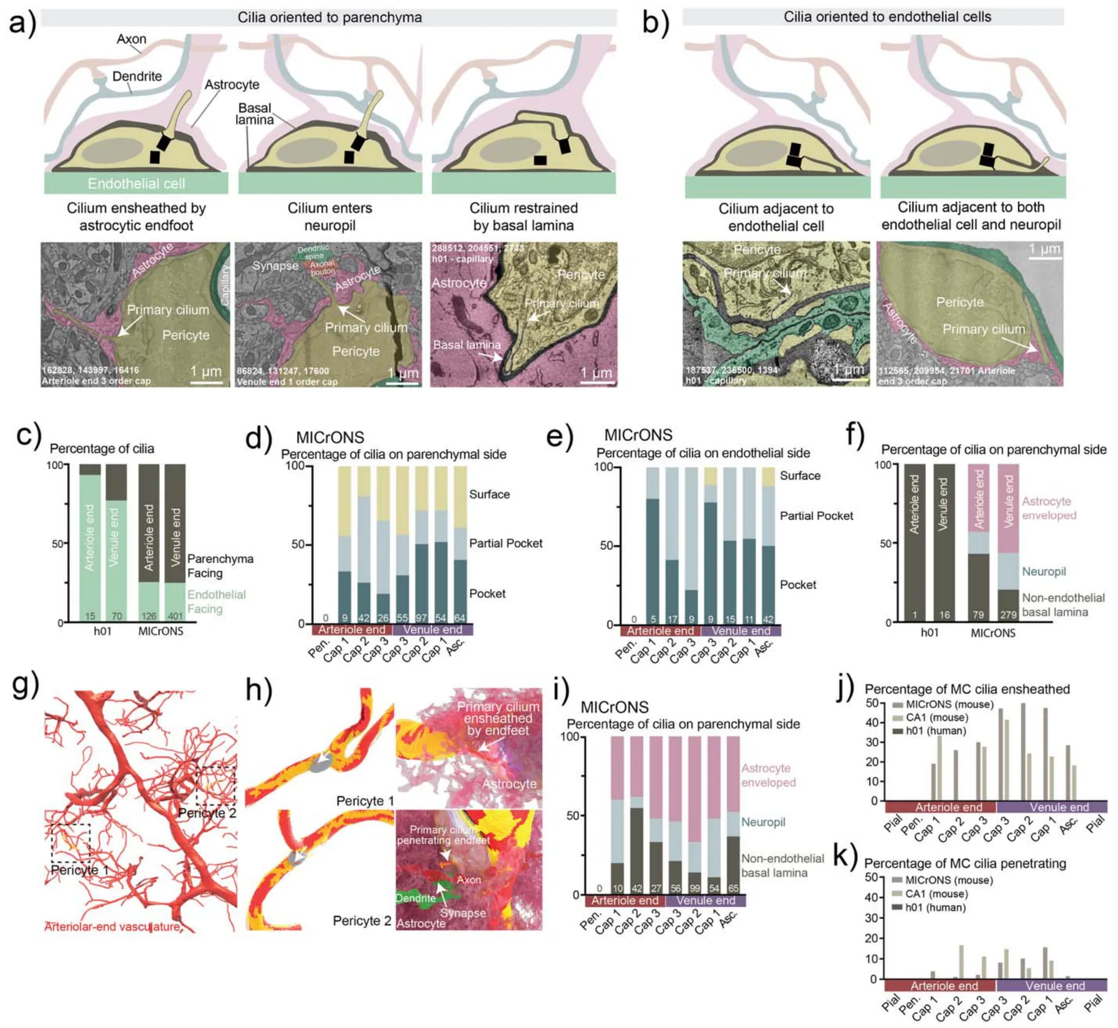
A primary cilium is a single antenna a cell puts out to feel its surroundings. Whether the cells of the brain's blood vessels have one, and where, had never been surveyed — because you need a volume to see a structure that is one micrometre long and attached to a cell you must first identify. Three volumes were read: MICrONS mouse visual cortex, a mouse hippocampal CA1 volume and the human h01 temporal cortex, plus nine other tissues.
What we found
Human cortical endothelial cells carry primary cilia, protruding into the lumen. Not one mouse endothelial cell in either mouse volume did — a species difference, not a sampling accident.
Mural cell cilia follow a gradient along the vascular tree: 83% of pial venular and 92% of ascending venular mural cells have one, against 3% of pial arteriolar and under 1% of penetrating arteriolar.
Three ways of building the base were found, including a partial pocket — some distal appendages at the plasma membrane and the rest on an invaginating one — in 25–50% of mural cells. Photoreceptors are the only other cell known to do this.
In mouse, about 75% of mural cell cilia point away from the vessel; most are ensheathed by an astrocyte endfoot, with the tip pressed into the astrocyte, and a few reach into the neuropil and pass close to synapses. In human, nearly all stay inside the basal lamina against the endothelium.
Endothelial centrioles sit consistently on the upstream side of the nucleus, against the direction of flow — which makes them a structural marker for which way the blood is going.
Figure 4 — where a mural cell points its cilium. Top left: one ensheathed by an astrocyte endfoot; next to it, one that enters the neuropil and runs past a synapse. The two human panels are h01 coordinates, in a different volume from the three below. Figure 4, Grubb, Chaddha, Lippincott-Schwartz, Ott & Mughal 2025, bioRxiv. Posted with all rights reserved; shown here by a co-corresponding author.
Go and look
162828, 143997, 16416Cilium ensheathed by an astrocyte endfoot — arteriole-end 3rd-order capillary (4a)
86824, 131247, 17600Cilium entering the neuropil past a synapse — venule-end 1st-order capillary (4a)
112565, 209954, 21701Cilium against both endothelium and neuropil — arteriole-end 3rd-order capillary (4b)
288512, 204551, 2743Cilium restrained by the basal lamina — human capillary (4a)h01 — a different volume, not resolvable here
187537, 236500, 1394Cilium against an endothelial cell — human capillary (4b)h01 — a different volume, not resolvable here
With Copenhagen and Boston · preprint
Distinct Ca²⁺ signatures of leptomeningeal fibroblast subgroups in awake mouse brains in health and inflammation
Chen He, Søren Grubb, Lechan Tao, Kıvılcım Kılıç, Xiao Zhang, Anna Devor, Neha Zahoor Sheikh & Changsi Cai
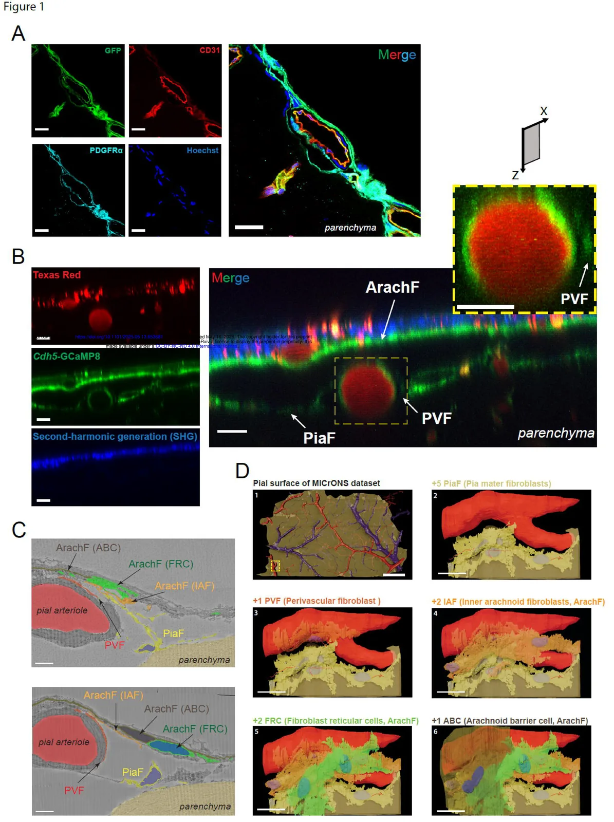
The leptomeninges are the two thin sheets between skull and cortex, built mostly of fibroblasts. Two-photon imaging in awake mice can watch them signal but cannot tell the groups apart with any confidence; the ultrastructure can. The pial surface is not segmented in the public MICrONS release, so this half of the paper is a hand-traced reconstruction: a CloudVolume cut-out around one pial arteriole, segmented cell by cell in TrakEM2.
What we found
Five kinds of fibroblast, told apart by position and ultrastructure: arachnoid barrier cells, fibroblast reticular cells and inner arachnoid fibroblasts in the arachnoid mater, pia mater fibroblasts on the surface, and perivascular fibroblasts wrapped around the pial vessels.
Each group has its own Ca²⁺ profile at rest in awake mice, and pia mater fibroblasts carry the largest transients.
Vasodilation during locomotion is followed by a fibroblast Ca²⁺ rise that depends on TRPV4 — a channel opened by stretch, in the order you would expect if the vessel moves first.
Seven days after a systemic LPS challenge, spontaneous Ca²⁺ in pia mater fibroblasts falls, and macrophages have infiltrated every leptomeningeal layer.
Figure 1 — panels C and D are the volume EM half: the five fibroblast groups around a pial arteriole, then the same reconstruction built up one group at a time over the pial surface of the dataset. Traced in TrakEM2, rendered in Blender. Figure 1, He et al. 2025, bioRxiv. CC BY-NC-ND 4.0.
Go and look
87644, 91688, 8900The pial arteriole the cut-out was taken aroundminnie35 — no nucleus table out here
88674, 88433, 9644Arachnoid and pia around the same vesselminnie35 — no nucleus table out here
89967, 89599, 9450Perivascular fibroblast on the pial vesselminnie35 — no nucleus table out here
91713, 92739, 9066Macrophage in the subarachnoid space, against a fibroblastminnie35 — no nucleus table out here
Single author · preprint
Ultrastructure of the brain waste-clearance pathway
Where does the brain's waste actually leave? The argument is usually between lymphatic vessels and the arachnoid villi, and it is usually made with tracers. This is the same question asked of the ultrastructure instead: follow the cells that carry waste and see where they put it down. Observational, and meant to generate hypotheses rather than settle one.
What we found
Twenty-one penetrating arterioles in the volume carry a perivascular space, and its depth is nothing like constant: from about 35 µm on one of them to over 500 on another.
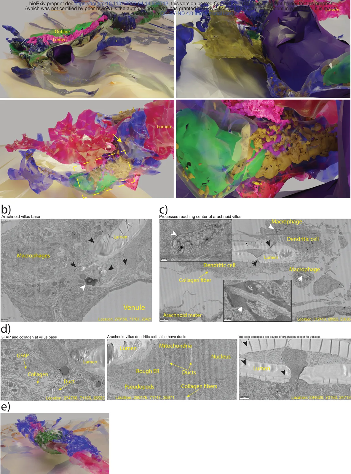
Meningeal macrophages carry indigestible waste in large lysosomes into the arachnoid villus and appear to exocytose it through ducts into the villus lumen, which drains to the venous sinus — a route a PEGylated tracer designed to avoid macrophages would never report.
Leptomeningeal fibroblasts reach macrophages with their primary cilia, which is a contact between the two cell types nobody was looking for.
Glia limitans astrocytes carry GFAP bundles 1–2 µm across that run from the brain surface to capillaries, and some reach the villus lumen itself.
Figure 3 — the arachnoid villus. Macrophages at the base (b), dendritic cell processes crossing the lumen (c), and the ducts through which lysosomal contents reach it (d). Figure 3, Grubb 2023, bioRxiv. CC BY-ND 4.0.
320752, 84727, 12738A perivascular space 508 µm deepminnie35 — no nucleus table out here
How this became the tools
Every study above was done the same way, and the way is slow. Find a cell. Decide what it
is from its ultrastructure and its shape. Write the coordinate down. Do it again, four
hundred times, and then count.
That loop is what the eight Jump tools automate. µJump is
the step where you stand at a coordinate and say what the cell is; the coordinate, not the
segmentation's nucleus id, is what identifies it — which is a rule these papers taught,
because around vessels the public segmentation is often wrong. The region-box download
writes the Colab notebook that cuts a block out of the volume and opens it in Blender,
which is the route the leptomeningeal reconstruction took by hand: a CloudVolume cut-out,
traced in TrakEM2 under FIJI, rendered in Blender.
The tools: µJump (MICrONS minnie65 and minnie35, where four of
these five studies were done), ηJump (the human h01 volume, the
comparison in two of them), and ωJump, which carries 62 further
volumes that nobody has published a soma list for.
Figure credits
Each figure is shown whole and downscaled for the web, never cropped to a single panel:
two of the five are published under a licence with a NoDerivatives term, and a cropped
panel is exactly what that term is about.
Vascular Biology 2023 — CC BY 4.0, reuse with attribution.
bioRxiv 2025.11.19.689283 — posted with all rights reserved, shown by a co-corresponding author.
The underlying imagery belongs to the consortia that released it, not to this site: see
Data & attribution. The text of this page is under
CC BY 4.0 and the
code behind it under the MIT License.